
Our Technology
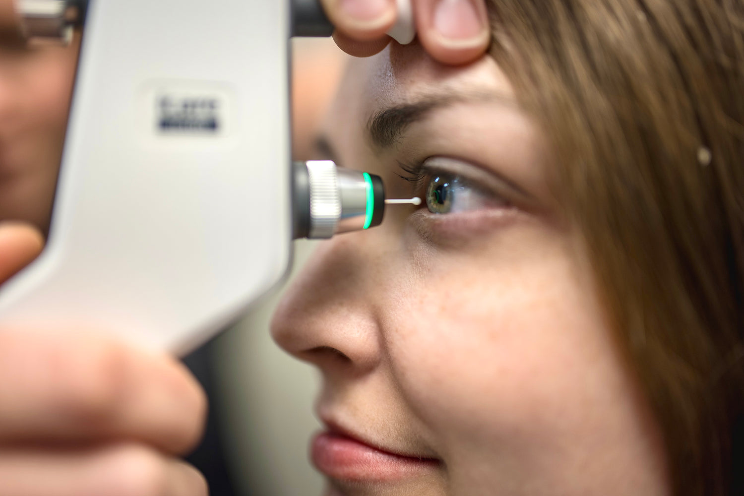
iCare Tonometry
A more comfortable way to measure eye pressure.
The comfort of all our patients is a priority, and we’ve heard many times that the dreaded “air puff” pressure check is most patients' least favourite part of having an eye exam… That’s why we upgraded years ago to the iCare Tonometer, a more comfortable way to measure eye pressure.
Elevated intraocular pressure (IOP) can be related to glaucoma or other diseases and is an important part of your comprehensive exam.
Benefits of the iCare tonometer include:
- No more puff of air! Quick and painless measurement that won’t make you jump out of your seat!
- Accurate and rapid testing that will collect six measurements and create an average for each eye
- Fully portable and handheld for increased accessibility
- No anesthetic drops needed
- Appropriate for all ages, including children.
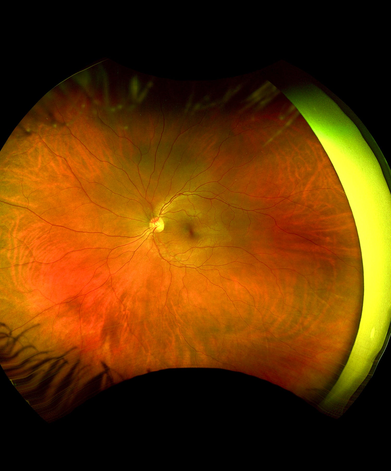
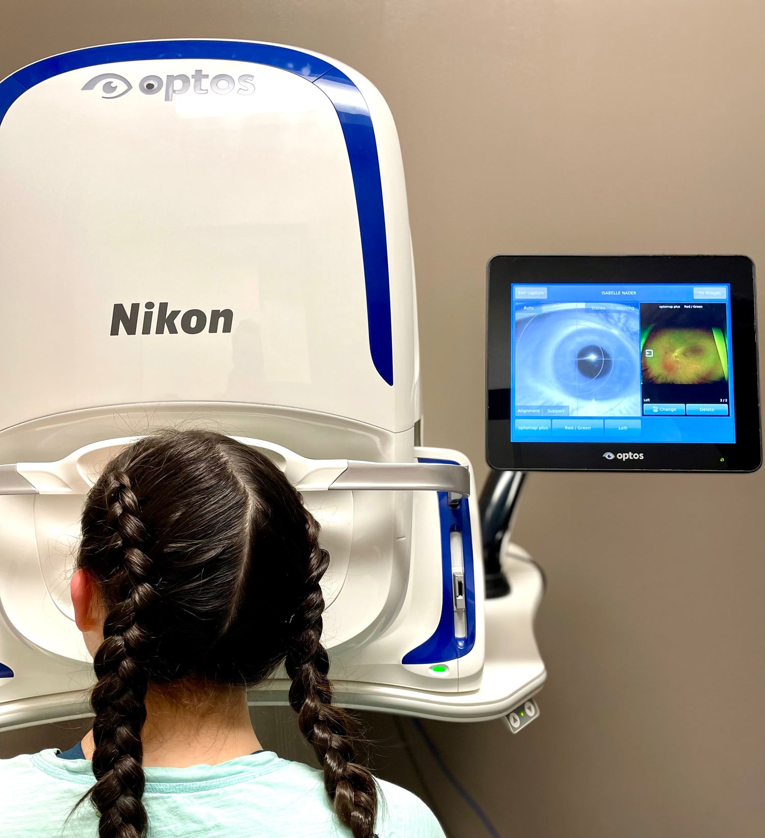
Optos Optomap Digital Retinal Imaging
A unique technology that captures a panoramic image of your retina in less than a second.
As an integral part of your eye examination, your eye doctor’s ability to complete a thorough retinal evaluation is enhanced by this amazing imaging technique.
Originally inspired to help capture images on children, this device is patient friendly and accessible to all ages. It is fast, easy, comfortable and safe.
While traditional retinal photos only show 15% of your retina at one time, we are able to capture more than 80% in a single high-resolution image! This will allow your doctor to quickly identify eye conditions such as retinal holes or detachments, macular degeneration, and glaucoma, as well as general health concerns, including diabetes, high blood pressure, or cardiovascular disorders.
Eye problems can develop without warning, progress with no symptoms, and lead to serious eye health issues and blindness. The optometrists at Insight Optometry recommend Optomap ultra-widefield retinal imaging for all patients, adults and children alike, even if you have no known health issues.
Optos Optomap Digital Retinal Imaging provides:
- In-depth viewing of the retinal layers (where disease can begin)
- Computerized image manipulation to enhance retinal assessment
- The ability to review your Optomap retinal image with your doctor during your examination
- A permanent clinical record which gives your doctor comparisons for tracking and diagnosing eye diseases at the earliest possible time
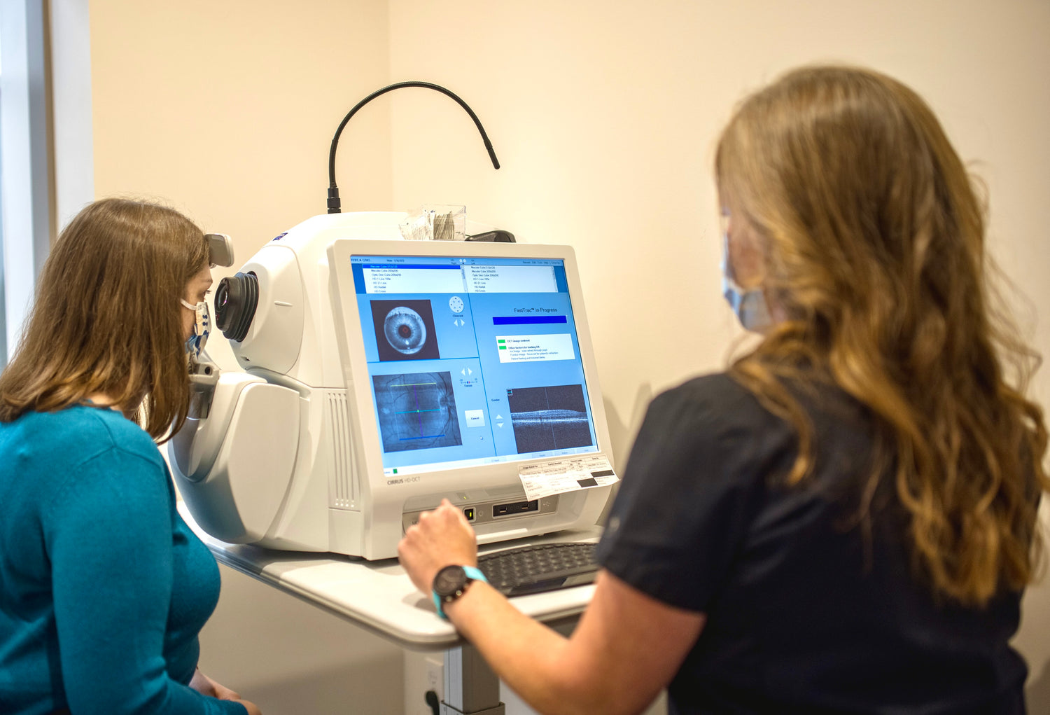
Zeiss Cirrus OCT (Optical Coherence Tomography)
Leading technology for advanced analysis of the eye's internal structures.
The Zeiss HD-OCT is a highly advanced instrument that will provide key information for the earliest detection and diagnosis of eye diseases.
As the world’s leading OCT innovator, ZEISS has been at the forefront of industry-defining advancements that have made OCT the standard of care. The OCT is an indispensable instrument used for screening, diagnosing and monitoring disease of the macula, optic nerve head, retina, and cornea.
Optic Nerve Analysis
Using Retinal Nerve Fiber Layer Analysis, your doctor can identify your risk factors for vision-threatening diseases, such as glaucoma and optic neuritis. The OCT will allow your doctor to monitor for microscopic changes, leading to a diagnosis of disease as early as possible, which is critical in protecting your sight. The OCT is the same technology utilized by glaucoma specialists (ophthalmologists).
Macular analysis
The macula is the most sensitive part of the retina and provides you with 20/20 vision and colour perception. The OCT gives a cross sectional view of this 0.3mm thick structure and allows for cellular analysis for early detection of retinal problems, such as age-related macular degeneration, diabetic retinopathy, macular holes, and side effects from medications.
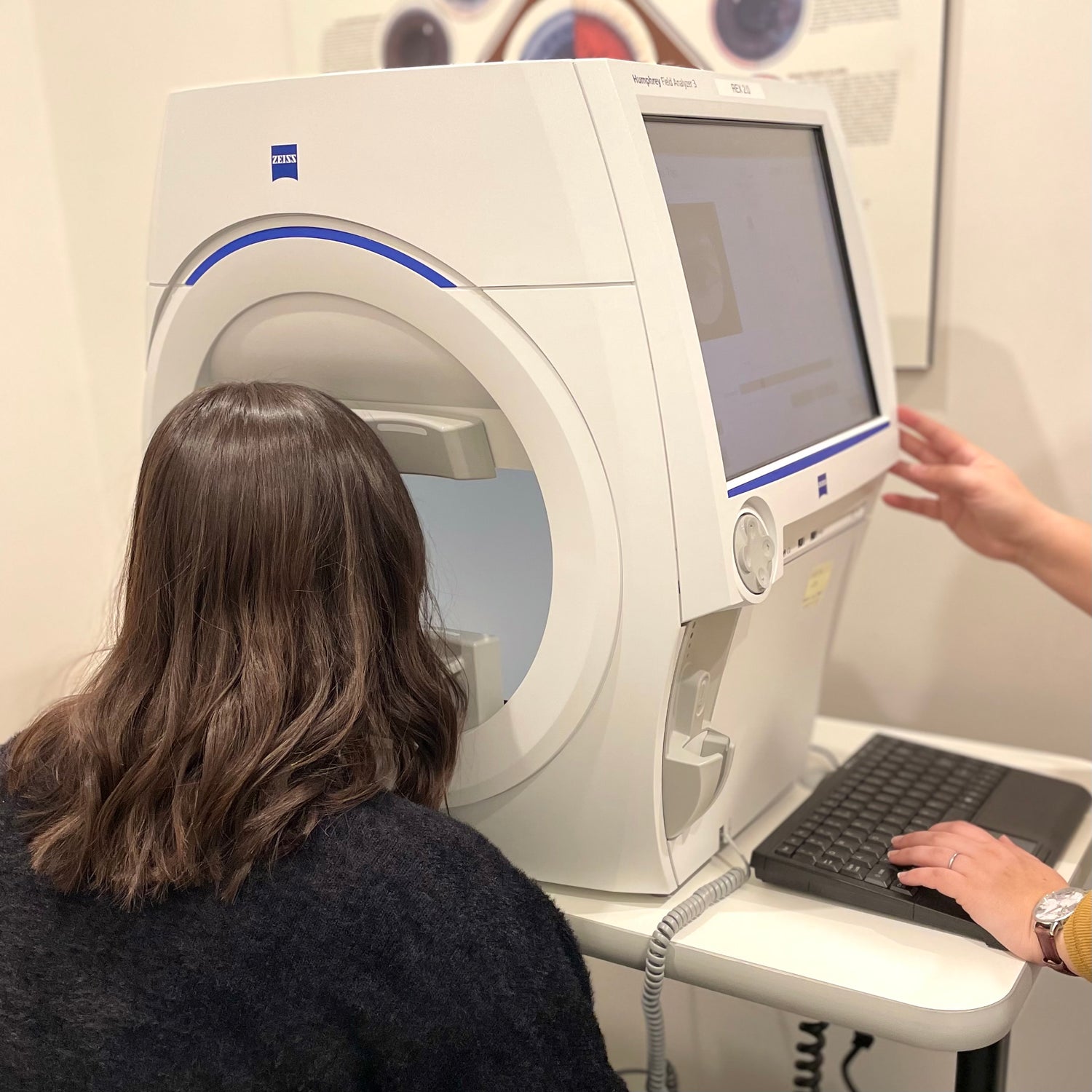
Humphrey Visual Field Analyzer 3
A vision test that maps out how you perceive the world around you.
The Humphrey Visual Field Analyzer 3 (HFA3) provides accurate, timely measurements of the entire visual field, including central vision (straight ahead) and side vision (peripheral).
The central area of the retina is the most sensitive to light, and we see best what is directly in front of us. The more peripheral areas of the retina are less sensitive, but they allow us to see, though less clearly, objects off to the side or above or below. Just how much we can see of the world around us is known as our visual field.
Through your feedback, the HFA3 maps out your entire visual field and sets the standard for visual field testing. Its advanced technology incorporates a liquid lens that automatically loads your most recent prescription in front of your eye for the test.
Your optometrist can utilize visual field information to assist in early diagnosis or monitoring of many disease processes, including glaucoma, brain tumours, neurological disorders, retinal problems, stroke, and many other conditions.
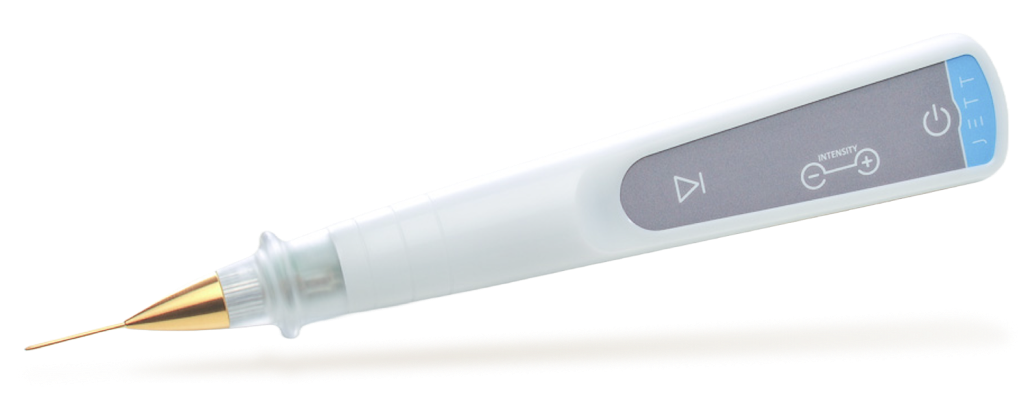
Jett Plasma Pen
A non-invasive treatment technology designed to support healthier, more comfortable eyes. Using controlled plasma energy, it targets underlying causes of irritation while protecting the delicate areas around the eyes.
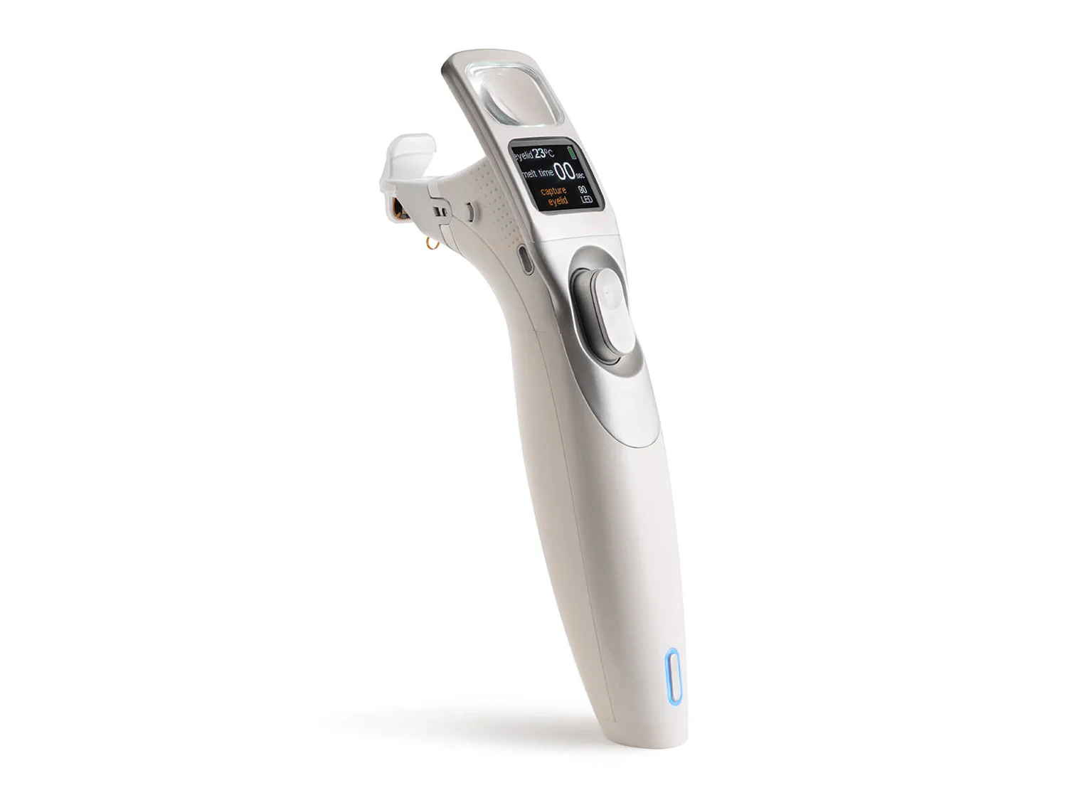
iLux
An in-office treatment that gently warms and expresses the meibomian glands to help restore healthy tear function. Designed for comfort and precision, it addresses one of the leading causes of dry eye.
Book an Appointment
Please note, not all optometrists are available through online booking - for more details, please call or email.